
 ×
×
The Short Answer: Auditory evoked potentials (AEPs) are small electrical signals recorded from the nervous system in response to auditory stimuli. Clinicians use these signals to assess hearing and auditory function at different points along the auditory pathway, from the cochlea to the auditory cortex.
AEPs give audiologists and other healthcare providers an objective window into the auditory system. An evoked potential test does not rely on a patient’s conscious response, which makes it useful when testing infants, young children, or any patient who cannot participate in a standard hearing test. Clinical applications range from newborn hearing screening to cochlear implant candidacy and the diagnosis of auditory neuropathy spectrum disorder. This guide covers what AEPs are, the main types used in clinical practice, how potential testing works in the clinic, and why these recordings matter for patient care.
When sound reaches the ear, it triggers a chain of electrical activity through the auditory system. The cochlea converts each auditory stimulus into neural impulses that travel up the auditory nerve, through the cochlear nucleus and brainstem, and finally reach the primary auditory cortex.
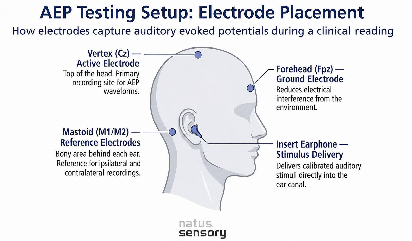
AEPs capture this electrical activity using surface electrodes placed on the scalp. Each signal is tiny, often less than a microvolt, so the recording system averages hundreds or thousands of responses to separate the brain’s response from background noise and unrelated EEG signal activity.
An evoked potential reflects sensory pathway activity. An event-related potential reflects cognitive processing. AEPs sit within the broader category of evoked potentials and share methods with peripheral nerve and spinal cord studies used elsewhere in neurology.
Each relay point along the auditory pathway produces measurable electrical activity:
Peak latencies and waveform shape shift with patient age, stimulus intensity, and the condition of the auditory system. Clinicians compare recorded waveforms against normative data to identify abnormalities.
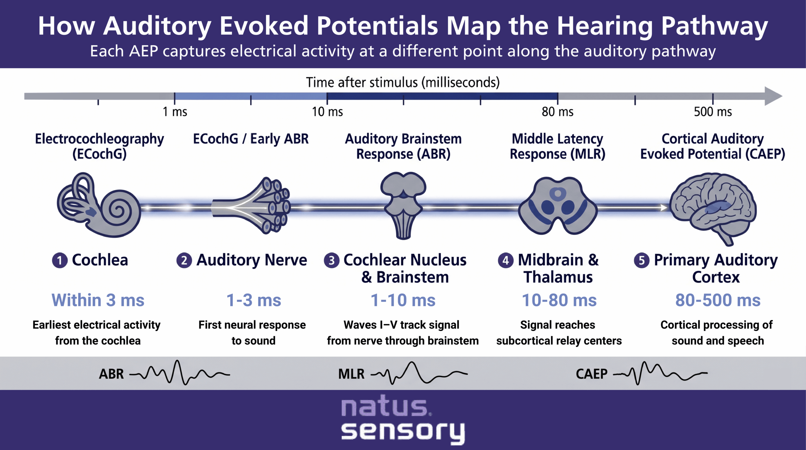
AEPs are grouped by when they occur after the stimulus. Each type tells a different story about auditory function.
Auditory Brainstem Response (ABR)
The auditory brainstem response is the most widely used AEP in clinical practice. Occurring within 1 to 10 milliseconds of the stimulus, ABR tracks electrical signals through the auditory nerve and brainstem, producing a series of waves (I through V) with predictable peak latencies in normal-hearing individuals. ABR forms the basis of most newborn hearing screening programs and remains the standard for identifying auditory neuropathy spectrum disorder.
Middle Latency Response (MLR)
The middle latency response occurs between 10 and 50 milliseconds after an auditory stimulus. It reflects activity in the thalamus and early cortex, bridging the brainstem and cortical levels of the auditory pathway.
Cortical Auditory Evoked Potential (CAEP)
CAEPs are recorded between 80 and 300 milliseconds after stimulation. They often show larger amplitude waves and are useful for evaluating cortical responses to speech stimuli, hearing aid benefit, and cochlear implant outcomes. CAEPs also give clinicians insight into auditory perception at higher levels of processing.
Auditory Steady State Response (ASSR)
ASSR uses modulated tones to evoke responses at the steady state of stimulation. It lets clinicians estimate frequency-specific hearing thresholds across a wide range of hearing loss severities, including severe-to-profound cases where ABR may reach equipment limits.
Electrocochleography (ECochG)
ECochG records the earliest electrical activity generated by the cochlea and auditory nerve, with a latency of approximately 1.5 ms (±0.2 ms) with the onset of the stimulus. The ECochG latency depends on which component you are measuring: the Cochlear Microphonic (CM), the Summating Potential (SP) and the Action Potential (AP), which is the most important clinically speaking. It supports the diagnosis of conditions such as Ménière’s disease/endolymphatic hydrops through it’s metrics SP/AP and is used to monitor auditory nerve function during intraoperative cochlear implant placement and acoustic neuroma surgery.
A clean recording depends on careful electrode placement, controlled stimulation, and a quiet test environment.
Typical setup:
Stimuli vary by test. Click stimuli are common for ABR because they activate a wide range of auditory nerve fibers. Tone bursts provide frequency-specific information. Speech stimuli are used when assessing cortical responses to real-world sound. Chirp stimuli are also used to compensate for the traveling-wave delay along the cochlea, causing neural activity from different frequency regions to arrive at the auditory nerve more synchronously.
For infants and young children, testing is typically done during natural sleep or light sedation to reduce movement artifact and improve signal clarity.
AEPs have become a foundational diagnostic tool across audiology, neurology, and neonatology. Common clinical applications include:
In cochlear implant candidates, AEP testing helps confirm that the auditory nerve can respond to an electrical pulse, which supports device selection and programming. After implantation, CAEPs can show whether cortical responses are developing as expected.
Each test in the audiologic battery has its role. AEPs complement behavioral audiometry and otoacoustic emissions testing rather than replacing them.
A common pattern: a newborn with present otoacoustic emissions and an absent auditory brainstem response likely has auditory neuropathy spectrum disorder. The combination of tests builds a clearer clinical picture than any single measure.
Clinicians compare waveform morphology, peak latencies, and inter-peak intervals against normative data. A significant difference from expected values, or a strong correlation coefficient between repeat recordings, supports confidence in the result.
Factors that affect interpretation include:
AEPs are sensitive to lesions at many points along the auditory pathway, but they do not replace imaging when structural diagnosis is needed. Results are interpreted alongside case history, otoscopy, tympanometry, and behavioral results when available.

Supporting Patient Care With AEP Testing
Auditory evoked potentials give clinicians an objective view of the auditory system at every level, from the auditory nerve to the primary auditory cortex. They support early identification of hearing loss, inform cochlear implant decisions, and confirm hearing status when behavioral audiometry is not an option. Paired with otoacoustic emissions and other measures, AEPs help build a complete clinical picture for infants, young children, and adults.
Natus Sensory builds auditory evoked potential systems designed with clinicians in mind, with the Bio-logic NavPRO ONE for AEP and addtional add-on licenses/modules like the OAE for testing across patient population. Explore our full hearing assessment and infant hearing screening portfolios, or contact our team to find the right fit for your clinic.
Sources:
053413 RevA