


 ×
×
Video otoscopy systems designed for high-resolution imaging of the ear canal and tympanic membrane, supporting accurate diagnosis, patient education, and hearing aid fitting workflows.
Products
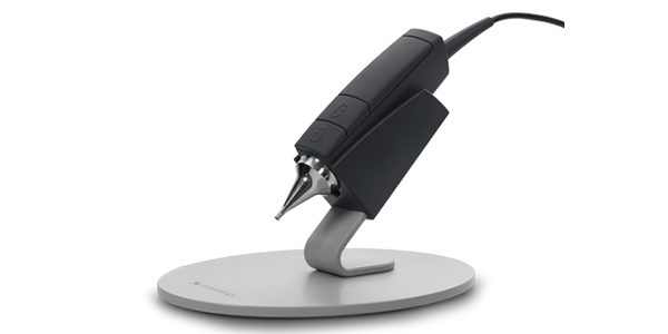
A digital video otoscope with improved image clarity, anti-fog imaging, USB plug-and-play setup, and seamless Otosuite® integration.
A video otoscope is a digital otoscope that uses a small video camera and powerful light source to display real-time, high-resolution images of the external ear canal and tympanic membrane on a screen. Unlike a traditional otoscope, which limits visualization to the clinician’s eye, a video otoscope allows the patient, family members, and care team to view the same image at the same time.
Clinicians use video otoscopy to inspect the eardrum, identify cerumen impaction prior to earwax removal, document tympanic membrane perforation and other ear problems, and support the diagnosis of conditions such as otitis media, acute otitis media, and bacterial infection of the ear canal. Stored images and otoscopic videos become part of the patient record and support patient education, client education, and clinical decision-making over time.
These capabilities support healthcare professionals in delivering accurate ear exams across a wide range of clinical applications, from cerumen assessment and middle ear visualization to patient counseling and hearing aid fitting workflows
Frequently asked questions
What is a video otoscope, and how is it different from a traditional otoscope?
A video otoscope is a digital otoscope that uses a video camera and light source to display real-time images of the ear canal and tympanic membrane on a screen. A traditional otoscope limits visualization to the clinician’s eye, while a video otoscope allows the patient, family, and care team to see the same image at the same time, supporting documentation, patient education, and shared decision-making. Learn more in our guide on Video Otoscopy vs. Standard Otoscopy for Clinicians.
What can clinicians see during video otoscopy?
Video otoscopy provides high-resolution views of the external ear canal and tympanic membrane. Clinicians use it to assess cerumen before earwax removal, document tympanic membrane perforation and identify foreign bodies.
How does the Aurical Otocam 300 integrate with hearing aid fitting workflows?
The Aurical Otocam 300 integrates directly with Otosuite® software and Aurical Freefit. The QuickView feature lets clinicians access the video otoscope from within the real ear measurement workflow to position the probe tube, verify mold placement, and document the procedure, all in a single software environment.
Does the video otoscope require batteries or charging?
No. The Aurical Otocam 300 is USB-powered and plug-and-play, eliminating the need to manage batteries or charging cycles between ear exams.
How does video otoscopy support patient and client education?
Showing patients real-time images of their own ear canal and tympanic membrane on a large screen helps them understand findings such as cerumen, ear infection, or tympanic membrane changes. Captured images can also be stored in the patient record and revisited during counseling about hearing loss, hearing aids, or follow-up care.
How are video otoscope images stored and shared?
Images and otoscopic video captured with the Aurical Otocam 300 are stored directly in Otosuite® software, where they become part of the patient record. Files can be exported and shared with other healthcare professionals as part of referral, documentation, or care coordination workflows.
Does Natus Sensory offer service and support for video otoscopes?
Yes. The Aurical Otocam 300 is a precision medical device that benefits from routine inspection and care to maintain image quality. Natus Sensory offers global service programs, technical service, and clinical education through training academies and online resources for hearing care professionals.

Request a quote
Ready to make a difference in patient care?
Learn more about our industry-leading solutions and how we can help advance your care capabilities.
053563 RevA