
 ×
×
3D ear scanning vs. ear impressions: which is better for audiologists?
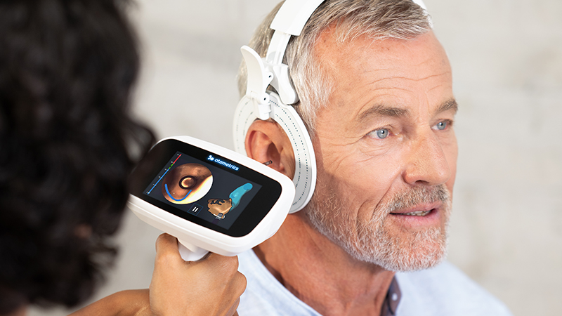
The Short Answer: 3D ear scanning captures the shape of the outer ear and ear canal as a digital file, while a traditional ear impression uses silicone material to create a physical impression that gets shipped to a lab. Digital ear scanning offers a faster turnaround, more comfortable, and reusable digital workflow for most audiology clinics, while traditional impressions continue to have a role in specific ear anatomies and clinical situations.
Custom earpieces depend on one thing above all else: an accurate record of the patient’s ear. For decades, that record was a silicone impression. Today, an ear scanner can digitally capture detailed ear anatomy without impression material entering the ear canal. Both methods provide the anatomical information needed to manufacture custom hearing aids, earmolds, and hearing protection. They differ in how the data is captured, how quickly it moves to the hearing aid manufacturer, and what happens when a device needs to be remade.
The National Institute on Deafness and Other Communication Disorders estimates that 28.8 million U.S. adults could benefit from hearing aids, and only about one in four adults 20 and over who could benefit has ever used them. While not every fitting requires a custom earpiece, every custom earpiece starts with an ear impression or digital scan.
3D ear scanning uses an optical scanner to map the geometry of the outer ear and ear canal, producing a digital impression that can be sent electronically to hearing aid manufacturers.
Two approaches exist in the market today:
Either way, the scanning process ends with a digital scan rather than a physical object.
The traditional impression process has changed very little in fifty years:
The method is familiar, inexpensive to start, and works on nearly every ear. Its weaknesses show up in the details: material blow-by past the otoblock, voids, distortion during removal, shipping delays, and shrinkage over time.
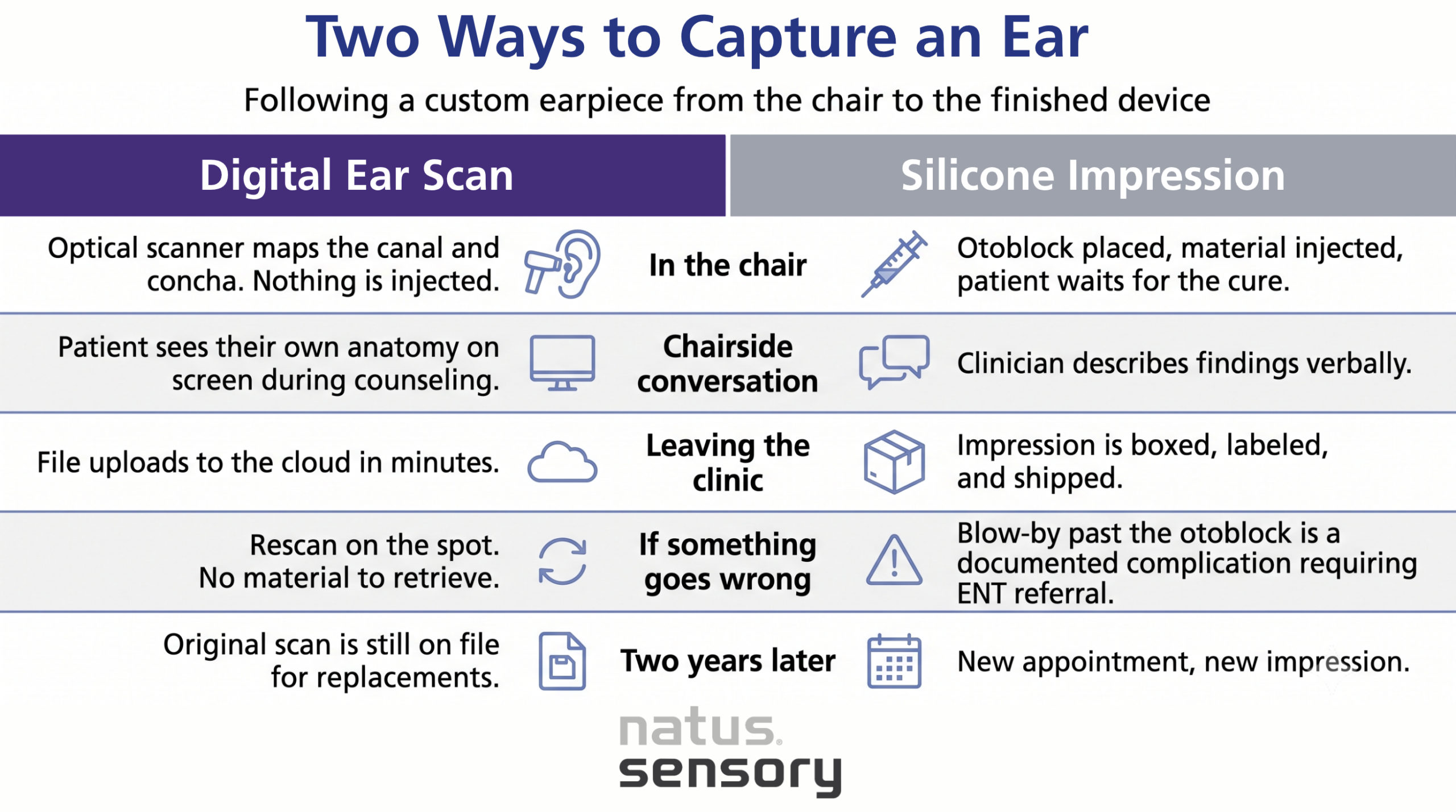
Both methods end with a custom earpiece. The key differences lie in how the data is captured, transferred, and managed afterward.
Capture and output
Getting the data to the manufacturer
What happens on reorders
Risk of distortion
Accuracy and fit
Accuracy is the argument audiologists care about most, and it comes down to remakes. Every remake means a second appointment, a longer wait for the patient, and clinic time that cannot be billed.
Natus Sensory reports that Otoscan reduces remake rates by 33 to 50 percent compared with the market average for custom products. A digital scan is not subject to the physical deformation or shrinkage that can affect impression materials after capture.
Traditional impressions can be uncomfortable for some patients. The impression material can feel cold and create a blocked sensation in the ear canal, while removal may be uncomfortable, particularly for patients with sensitive ears.
There is a clinical safety dimension as well. A case series published in the Annals of Otology, Rhinology & Laryngology documented six patients in whom impression material entered the middle ear during earmold fitting. The authors noted that these cases require ENT referral, that blind in-office removal carries real risk when the tympanic membrane cannot be fully examined, and that no standardized management approach exists once material has passed the otoblock. Related case reports have described silicone discovered in the middle ear years after the original fitting.
Digital ear scanning eliminates several challenges associated with traditional impressions, including the risk of material extending past the otoblock and the need for impression curing time. Patients can see their own ear scan in real time on screen. Showing a patient the 3D model of their own ear canal gives the audiologist a visual aid during hearing aid selection, and it pairs naturally with the counseling conversation that already happens during hearing aid fitting.
A silicone impression has to be boxed, labeled, and shipped. Electronic transmission can allow the manufacturer or lab to begin processing sooner than with shipped physical impressions.
Stored scan data also pays off later. The same stored scan may be submitted to different participating manufacturers for compatible custom products.
Custom earpieces are not limited to amplification. Regardless of protector type, NIOSH recommends individual fit testing to confirm adequate attenuation. Stored ear scans can simplify replacement orders when the existing scan remains clinically appropriate and is accepted by the manufacturer.
A pilot study published in Bioengineering compared the two techniques directly using the Natus Otoscan. Adult earmold users wore earmolds made from each method, one set per week in randomized order.
The authors concluded that 3D ear scanning is a viable consideration for audiology practices.
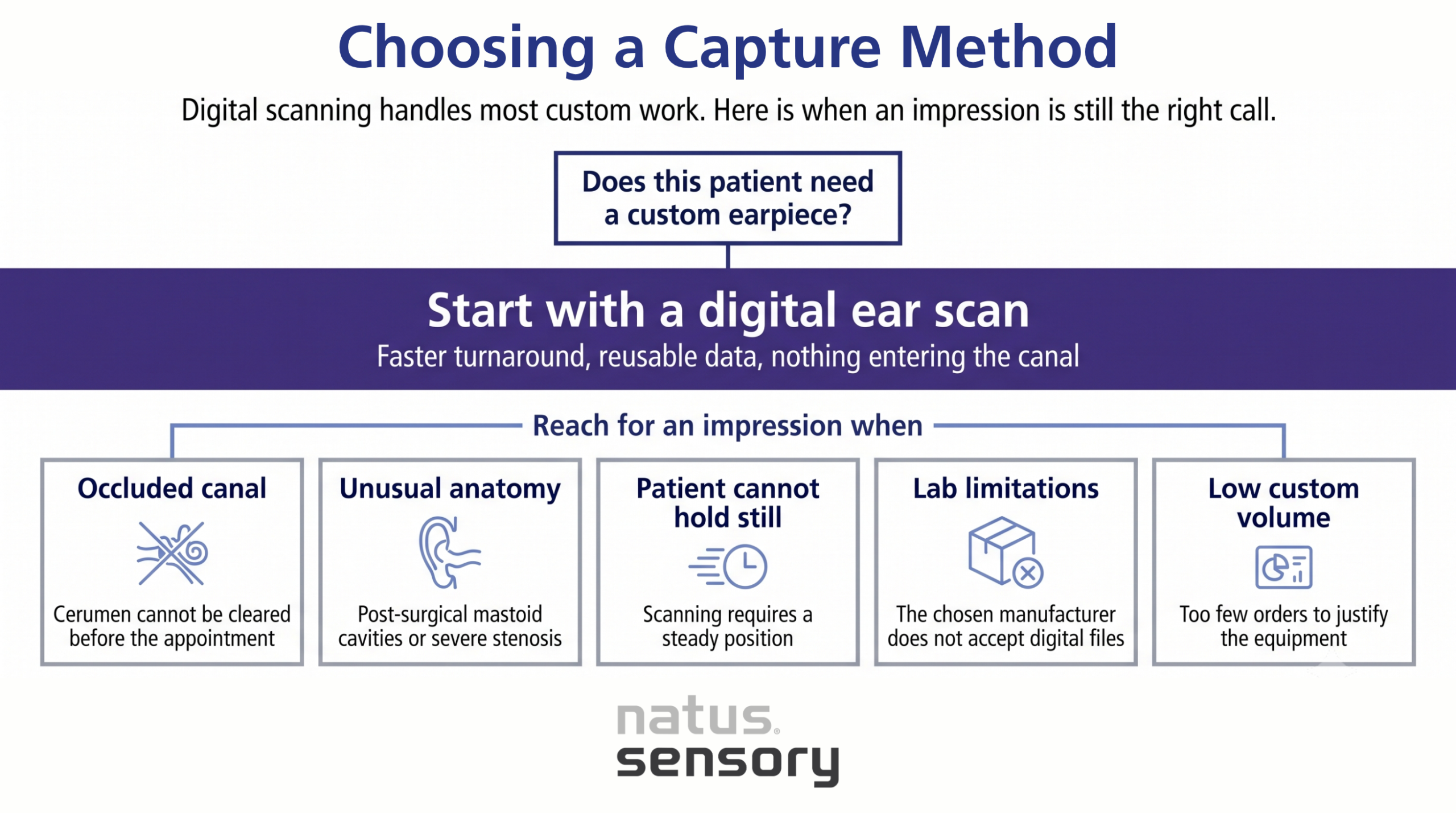
An experienced audiologist keeps both options available. Traditional ear impressions remain the better choice when:
An ear scanner is a capital purchase, so the math depends on volume. Higher-volume clinics may offset the capital cost through reductions in remakes, shipping, and workflow time, depending on local economics and usage.
Scanning is a clinical skill that improves with repetition, and structured training shortens the ramp. Training multiple qualified staff members in scanning can increase scheduling flexibility, subject to local scope-of-practice requirements.

Choosing the right approach for your clinic
For clinics with regular custom-device volume, 3D scanning may offer meaningful workflow, patient-experience, and data-management advantages. Published research supports it as a viable alternative to traditional impressions for patients who can be scanned successfully. Traditional silicone impressions still deserve a place in the drawer for difficult anatomy and edge cases.
Many clinics may choose to incorporate scanning as a routine option while maintaining competency in traditional impressions for cases where they remain appropriate.
Otoscan from Natus Sensory captures over one million data points per ear and supports digital scan uploads through Otocloud for electronic submission to participating manufacturers, with in-person and on-demand training included. To talk through how digital ear scanning would fit your clinic’s workflow, contact our team.
Sources:
054610 RevA