
 ×
×
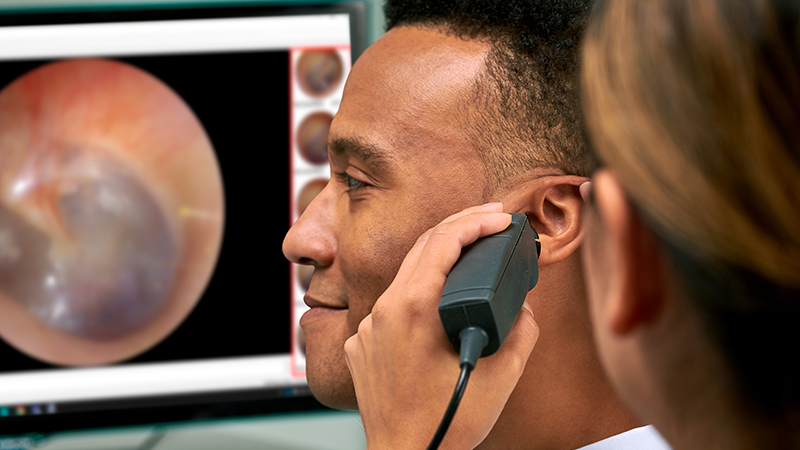
Video Otoscopy: When it’s worth using over standard otoscopy in the clinic
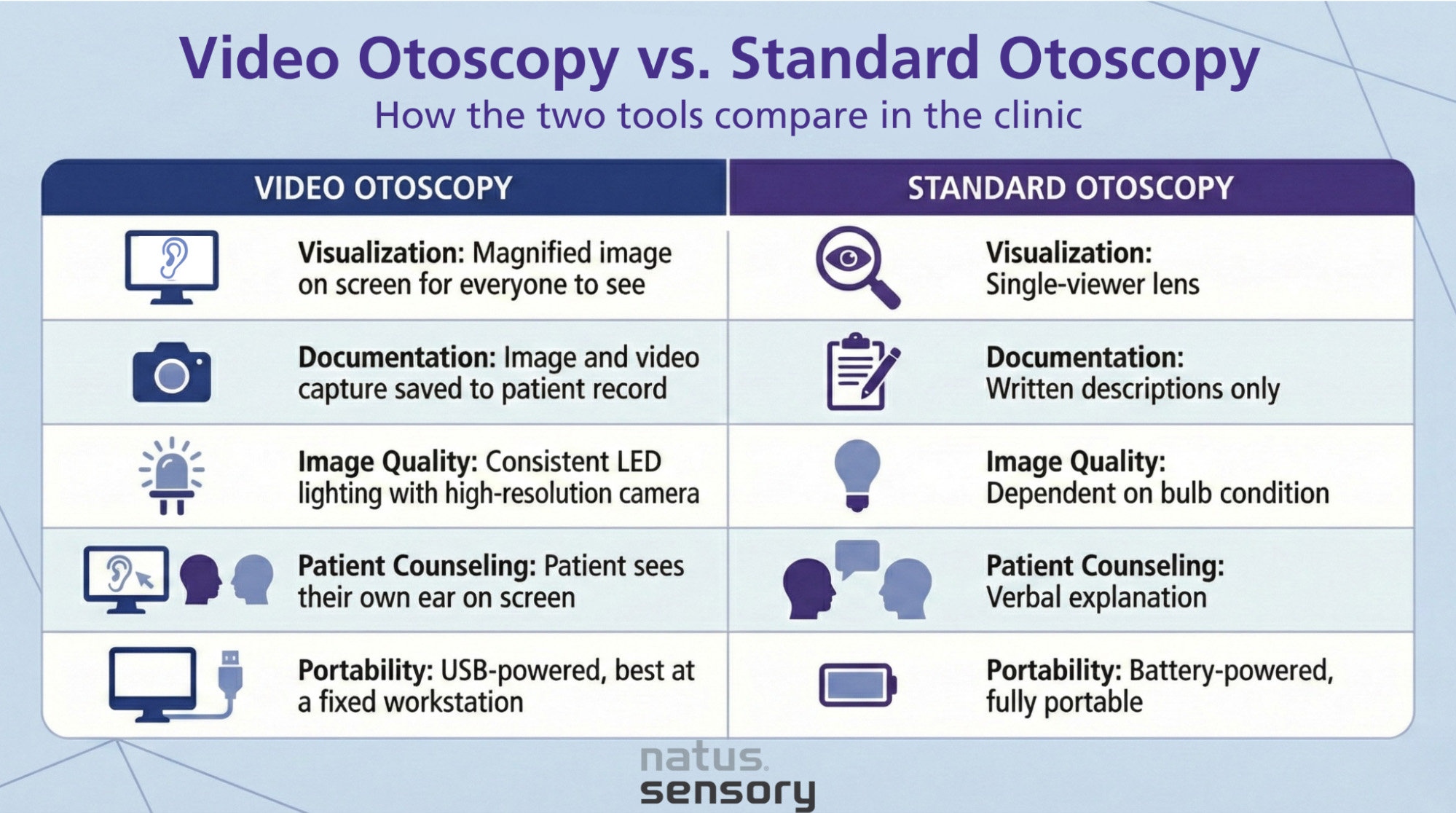
At a glance: Video otoscopy is worth using when you need magnified, shareable views of the ear canal and tympanic membrane that a standard otoscope can’t provide. It offers higher image quality, real-time visualization on a screen, and the ability to capture images for the patient record, making it especially useful for diagnosing ear infections, monitoring chronic ear conditions, and counseling patients.
Standard otoscopy remains reliable for quick assessments and routine exams. But as documentation requirements grow and clinics look for ways to improve diagnostic confidence, many healthcare professionals are finding that a video otoscope fills gaps traditional devices leave open.
Video otoscopy is an otoscopic examination that uses a camera integrated into the otoscope head. The camera feeds a magnified, real-time image of the external auditory canal and eardrum to a screen, letting the clinician, patient, and care team view the ear structures at the same time.
A traditional otoscope uses a light source and magnifying lens, limiting the view to one person looking through a small eyepiece. A video otoscope replaces that lens with a camera sensor, often paired with brighter LED illumination. The result is higher magnification, better color accuracy, and the ability to capture still images or video recording for the medical record.
Diagnosing ear infections and Otitis Media
Otitis media affects roughly 740 million people worldwide each year, and by age 10, about 90% of children will have experienced at least one episode of middle ear effusion. Distinguishing between acute otitis media, otitis media with effusion, and otitis externa depends on accurately visualizing the tympanic membrane. The appearance, position, and color of the eardrum all matter for an accurate diagnosis.
Research published by the AAFP found that roughly one-third of otoscopes used in private practices had suboptimal light output, contributing to misdiagnosis. Video otoscopes address this with brighter LED lighting and digital magnification that makes subtle tympanic membrane changes easier to spot.
Monitoring chronic ear infections and ear disease
For patients with chronic ear infections or recurring ear disease, capturing otoscopic images at each visit creates a visual timeline in the patient record. This is particularly useful for tracking tympanic membrane perforation, chronic infection, or retraction pockets before referring for surgical procedures. A general practitioner can document findings and share them with an ENT specialist without requiring an additional in-person visit.
Foreign body identification
Identifying a foreign body or object in the ear canal is another scenario where video otoscopy has a clear advantage. The magnified screen view helps clinicians pinpoint the exact location of the object and guide removal instruments in real time, reducing the risk of pushing it deeper or damaging the external ear canal or tympanic membrane.
Guided ear cleaning and deep ear flush
When cerumen is deeply impacted, performing a deep ear cleaning or deep ear flush under video otoscopy guidance allows the clinician to see exactly what they’re doing throughout the process. This reduces the chance of accidental trauma to the external ear canal or tympanic membrane.
Patient education and counseling
When patients see their own ear canal and tympanic membrane on a screen, they better understand their condition and why a particular treatment is recommended. This shared visual experience makes it easier for clinicians to explain findings like fluid behind the eardrum, signs of chronic infection, or the need for a referral. It also supports more productive conversations around treatment compliance and follow-up care.
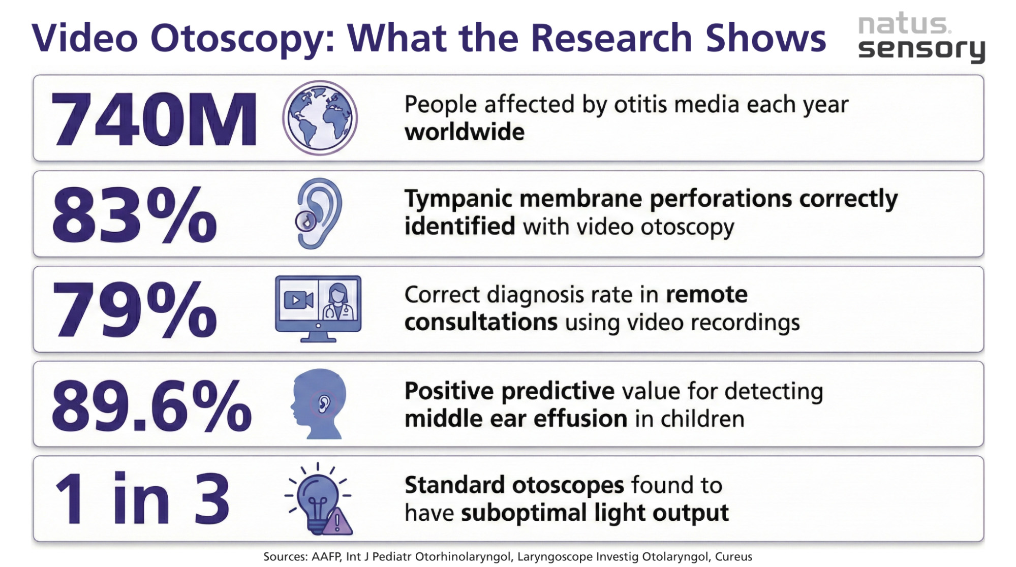
Multiple studies have compared video otoscopy to standard otoscopy. The results consistently show comparable or improved diagnostic accuracy:
Standard otoscopy remains practical and appropriate in certain situations:
Many clinics use both tools, reaching for the video otoscope when the situation calls for better visualization, documentation, or patient communication.
Health care professionals like audiologists use video otoscopy for pre-assessment screening before hearing tests and for documenting ear canal conditions before hearing aid fitting or digital ear scanning. ENTs use it to document ear disease and counsel patients before surgical procedures. General practitioners benefit from the improved visualization, especially when sharing findings with a specialist remotely.
It’s also a strong training tool. Because the image displays on a screen, supervisors can observe and guide trainees during an otoscopic examination without repeating the exam. A previous study in Advances in Medical Education and Practice found that video otoscopes improved agreement between trainees and supervisors and reduced repeated confirmatory exams.
Video otoscopy gives a detailed surface-level view, but it cannot see beyond the eardrum into the inner ear or provide a transverse slice of deeper structures like a CT scan can. If you suspect cholesteatoma extending beyond the tympanic membrane, an auditory canal mass, or complications involving the inner ear, advanced imaging is the appropriate next step. Video otoscopy and CT serve complementary roles.
Choosing the right tool for the exam
Video otoscopy earns its place in the clinic when you need better visualization, when documentation matters, or when showing a patient their ear structures will improve understanding and compliance. Research shows it supports diagnostic accuracy at a level comparable to or better than standard otoscopy, with added capabilities like image capture, video recording, and remote consultation.
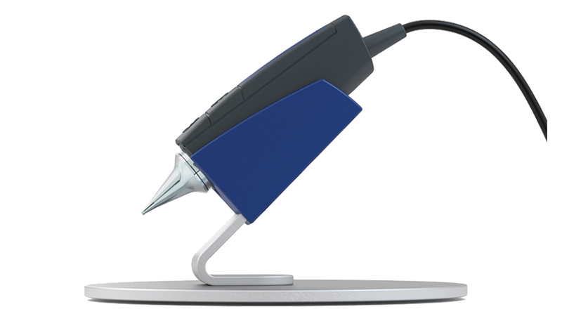
Natus Sensory’s Aurical® Otocam 300 Video Otoscope is designed with these clinical needs in mind, featuring high-resolution imaging, direct software integration, built-in condensation control, and USB power for a clean workspace.
Ready to see how video otoscopy fits into your practice? Contact Natus Sensory to learn more about the Aurical® Otocam 300 and explore solutions for your clinic.
Sources:
053104 RevA