
 ×
×
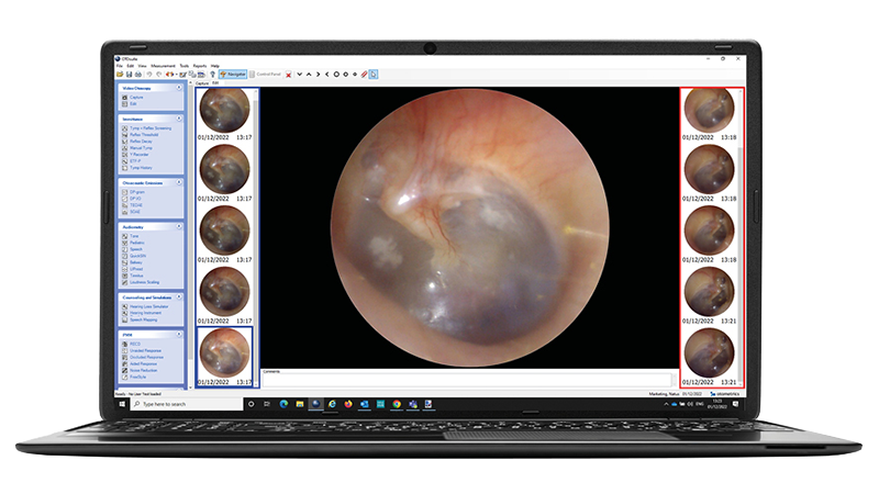
Otoscope With Camera: A Clinical Buying Guide
The Short Answer: In clinical settings, the top-performing otoscope with a camera is a video otoscope that combines high-resolution imaging, a bright LED light source, and real-time image capture. These tools give healthcare professionals a clear view of the ear canal and tympanic membrane. For home use, consumer-grade ear camera devices have improved significantly but still fall short of clinical-grade diagnostic accuracy.
A standard otoscope uses a light source and a magnifying lens to help a healthcare provider look into the outer ear. A video otoscope or digital otoscope adds a small digital camera to the setup, capturing live images or video of the ear canal and eardrum.
This matters clinically because:
Research supports the clinical value here. A 2017 study published in the International Journal of Pediatric Otorhinolaryngology found that video-otoscopy collected by a healthcare facilitator achieved diagnostic accuracy comparable to traditional otoscopy performed by a general practitioner, a finding that strengthens the case for digital tools in distributed care settings.
These three terms get used interchangeably, but they are not the same device.
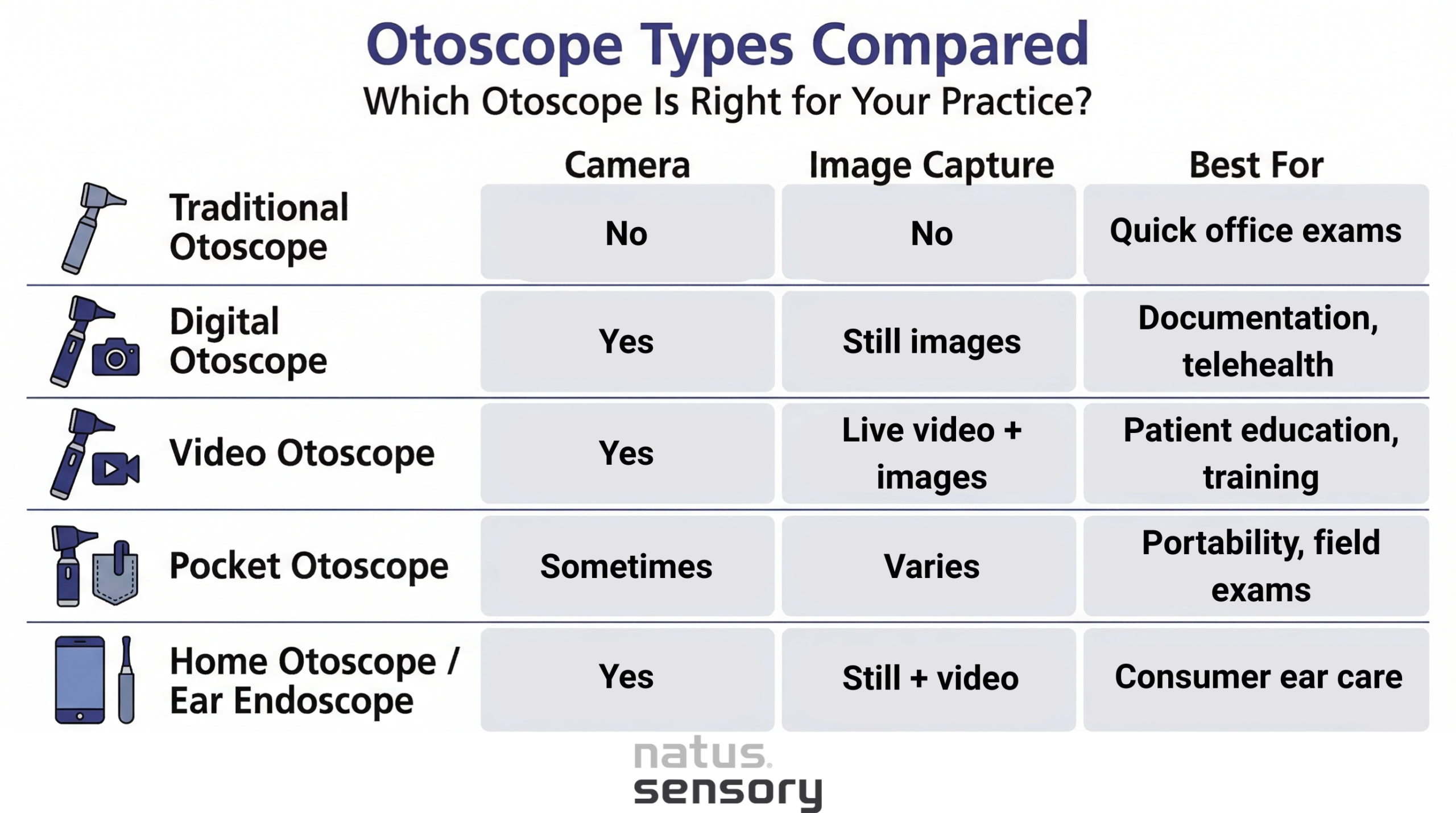
For clinical environments, a video otoscope is generally the most functional. It supports workflows across audiology, ENT, primary care, and pediatrics.
Image and Video Quality
The digital camera resolution determines how clearly you can see the tympanic membrane, spot ear wax buildup, identify foreign bodies, or detect early signs of an ear infection. Look for at least 1080p resolution in clinical-grade devices.
Light Source
A bright, adjustable LED light is one of the most important parts of any otoscope. It illuminates the ear canal without generating excessive heat. Weaker light sources make it harder to see fine details of the eardrum.
Magnification
The magnifying lens or x magnification level affects how much detail is visible. Clinical video otoscopes typically offer 3x to 10x magnification. Higher magnification helps with identifying subtle changes in the tympanic membrane.
Battery Type and Life
Rechargeable battery: Standard in premium and clinical-grade otoscopes. Better for high-volume clinical use.
Alkaline battery: Common in pocket otoscopes and lower-cost home otoscope models. Easy to swap, but adds ongoing cost.
Connectivity
Many digital otoscope models connect to a monitor, tablet, or computer. Connection options include a USB cable to a PC or laptop, wireless Bluetooth pairing, or direct screen output to a wall-mounted display. This is especially useful in audiology and ENT clinics where real-time patient viewing is part of the workflow.
Portability
A pocket otoscope with a pocket clip works well for rounds or home visits. Larger video otoscope systems suit in-office exam rooms with fixed monitor setups better.
The gap between a clinical-grade video otoscope and a consumer ear camera tool is larger than most people expect.
| Clinical (Professional Use) | Home Use |
Healthcare professionals and medical professionals need:
|
Consumer home otoscopes and ear endoscope products have grown into a real market. Parents check their children’s ears, adults monitor known ear conditions, and some people use them as an ear cleaning aid. For home use, reasonable priorities include:
These devices are not medical devices cleared for diagnostic use in most cases. Findings should always be followed up with a healthcare provider for accurate interpretation. |
A 2024 study in Laryngoscope Investigative Otolaryngology found digital otoscopy to be a practical tool for remote consultations, with strong clinician acceptance, reinforcing that image quality and connectivity are not optional features for professional-grade devices. A systematic review published in European Archives of Oto-Rhino-Laryngology further confirmed that video otoscopy over the past decade has shown reliable diagnostic performance across a range of otological conditions.
Knowing what you are seeing on-screen makes any ear camera more useful.
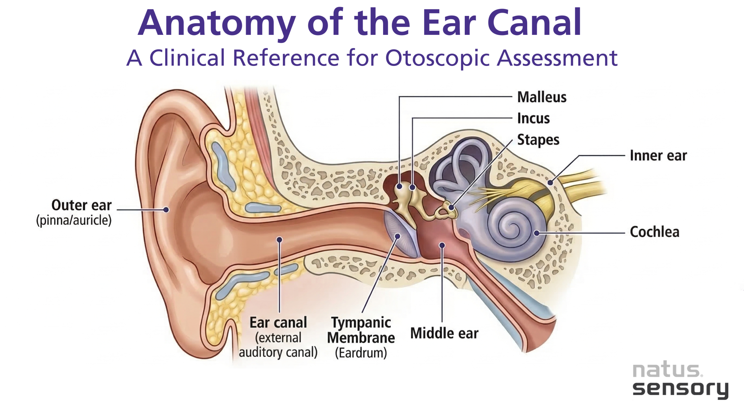
A healthy tympanic membrane appears pearly gray with a visible light reflex. Redness, bulging, fluid, or perforation can indicate an ear infection, fluid in the middle ear, or other conditions requiring treatment. Research published in Cureus (2024) found that video otoscopy can accurately predict middle ear effusion in children when compared to tympanometry, supporting its role in earlier detection in pediatric care.
Some consumers use ear camera tools primarily for ear wax removal and ear cleaning. It is worth understanding the limits here.
Tools often marketed together with ear cameras:
A note on cotton swabs: The American Academy of Otolaryngology advises against inserting a cotton swab into the ear canal. It can push wax deeper toward the eardrum, cause trauma, or introduce foreign bodies. If wax buildup is significant, professional wax removal or irrigation by a healthcare professional is the safer path.
Medical students typically start with a basic otoscope kit, but exposure to video and digital otoscope systems is increasing in clinical education.
Benefits of video otoscopy in training:
Early detection of conditions like otitis media or tympanic membrane perforation is easier to teach when the image is visible to both student and instructor simultaneously. A study in the Brazilian Journal of Otorhinolaryngology confirmed that video otoscopy shows strong diagnostic accuracy in detecting tympanic membrane perforations, findings that support its use in both clinical training and active patient care.
Choosing the Right Otoscope for Your Practice
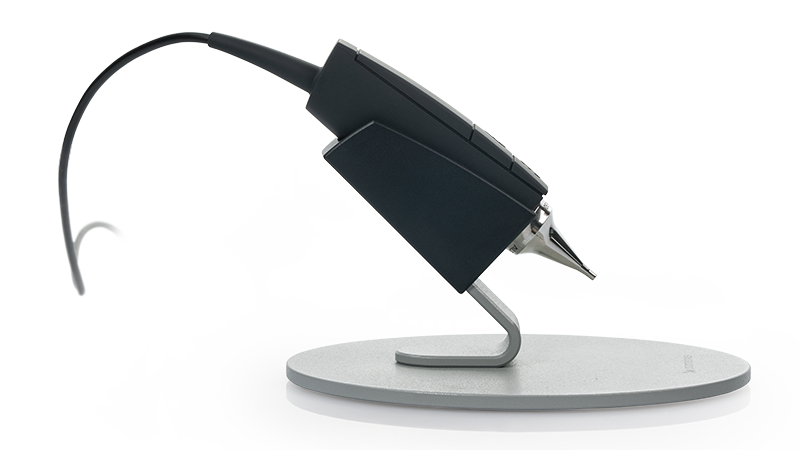
The best otoscope with a camera depends on how you plan to use it. For healthcare professionals, a clinical-grade video otoscope with a high-resolution digital camera, a reliable LED light source, and image documentation is the right tool. For home use, a consumer ear camera can offer general visibility, but findings should always be reviewed by a qualified healthcare provider.
At Natus Sensory, the Aurical Otocam 300 is built for clinical environments that require accurate visualization of the tympanic membrane and outer ear. If you work in audiology, ENT, or a related specialty and want to explore how video otoscopy fits your diagnostic workflow, connect with our team.
Sources:
053411 RevA